Spider-like cells inside the brain, spinal cord and eye hunt for invaders, capturing and then devouring them. These cells, called microglia, often play a beneficial role by helping to clear trash and protect the central nervous system against infection. But a new study by researchers at the National Eye Institute (NEI) shows that they also accelerate damage wrought by blinding eye disorders, such as retinitis pigmentosa.

Retina India is a not-for-profit organization, registered with the Charity Commissioner, Mumbai, India, established for empowering people with retinal disorders, and bringing them and their families on a common platform with physicians, researchers, counselors, low vision and mobility experts and other specialists.
Showing posts with label Treatment. Show all posts
Showing posts with label Treatment. Show all posts
Saturday, July 4, 2015


Thursday, April 23, 2015
Scleral thinning seen following intravitreal anti-VEGF injections
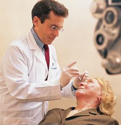
Research conducted at the University Hospital in Bern, Switzerland suggests that scleral changes may occur in patients following repeated intravitreal injections of anti-VEGF agents in the same quadrant.
Thursday, October 2, 2014
Stem cells close to cornea may help treat Macular Degeneration or Retinitis Pigmentosa
Scientists at the Southampton General Hospital and the University of Southampton have found a pool of stem cells around the corneal limbus that can be induced to become retinal cells. Such cells can then potentially be transplanted into the eye of the patient of macular degeneration or retinitis pigmentosa to stop the disease or even cure it.
Wednesday, September 17, 2014
Japanese woman is first recipient of next-generation iPS stem cells for macular degeneration
A Japanese woman in her 70s has become the first person in the world to receive retinal cells derived from induced pluripotent stem cells (iPS). In a two-hour procedure on September 12, 2014, a team of three eye specialists lead by Dr Yasuo Kurimoto of the Kobe City Medical Center General Hospital, Japan, implanted a 1.3 by 3.0 millimetre sheet made of retinal pigment epithelium (RPE) cells into one eye of this patient, who was diagnosed with age-related macular degeneration (AMD).
Wednesday, July 23, 2014
Treatment efficacy of topical unoprostone isopropyl in patients with retinitis pigmentosa
A study was conducted by Dr Akiyama & associates from the Department of Ophthalmology, Graduate School of Medical Sciences, Kyushu University, Fukuoka in Japan to evaluate the treatment effect of topical unoprostone isopropyl (unoprostone) (in the form of eye drops) on patients with retinitis pigmentosa (RP). This medication is currently used in glaucoma.
Forty patients with typical forms of RP were included in the study, out of which 17 patients were treated with 0.12% topical unoprostone twice daily in a randomly selected eye. The efficacy of the treatment was monitored by visual acuity and visual field testing using the Humphrey Field Analyzer (HFA). In addition, 12 RP patients who were included this study and 12 normal subjects were evaluated in terms of their macular blood flow of both eyes after instillation of unoprostone using the laser speckle method.
Forty patients with typical forms of RP were included in the study, out of which 17 patients were treated with 0.12% topical unoprostone twice daily in a randomly selected eye. The efficacy of the treatment was monitored by visual acuity and visual field testing using the Humphrey Field Analyzer (HFA). In addition, 12 RP patients who were included this study and 12 normal subjects were evaluated in terms of their macular blood flow of both eyes after instillation of unoprostone using the laser speckle method.
Is Valproic Acid effective in treating patients with Retinitis Pigmentosa?
There have been some reports in academic journals about the use of Valproic acid (VPA) in Retinitis Pigmentosa (RP). We thought we can review the current status of these trials and evaluate where we stand in the outcomes.
Saturday, April 27, 2013
Advanced Cell Technology initiates higher-dosage patient treatment in European clinical trial for Stargardt's Disease
Advanced Cell Technology, Inc., a pioneer in the field of stem cells for treating conditions such as macular degeneration. has announced treatment of the first patient in the third dosage cohort, and seventh patient overall, in its European Phase I clinical trial for Stargardt’s macular dystrophy (SMD) using retinal pigment epithelial (RPE) cells derived from human embryonic stem cells (hESCs).
The patient was injected with 150,000 hESC-derived RPE cells, as compared with the 100,000-cell dose used in patients of the second cohort. The surgery was performed on Friday, April 19, without any complications, and the patient is recovering uneventfully.
The company declared that they are past the halfway point in all three of their clinical trials on both continents. They have also announced that the European Medicines Agency's (EMA) Committee for Orphan Medicinal Products (COMP) has officially granted their hESC-derived RPE cells orphan medicinal product designation for the treatment of SMD. This will likely provide a number of benefits to them, as in protection from competition, as well as for the patients through reduced fees.
The Phase 1/2 trial is designed to determine the safety and tolerability of hESC-derived RPE cells following sub-retinal transplantation in patients with SMD at 12 months, the study’s primary endpoint. It will involve a total of 12 patients, with cohorts of three patients each in an ascending dosage format.
Stargardt’s disease or Stargardt’s Macular Dystrophy is a genetic disease that causes progressive vision loss, usually starting in children between 10 to 20 years of age. Eventually, blindness results from photoreceptor loss associated with degeneration in the pigmented layer of the retina, called the retinal pigment epithelium, which is the site of damage that the company believes the hESC-derived RPE may be able to target for repair after administration. Clink here to read more,
Saturday, March 23, 2013
A study for new treatments for wet age-related macular degeneration (AMD)
A University of Wisconsin School of Medicine and Public Health scientist, Dr Nader Sheibani, is spearheading a study of new treatments for wet age-related macular degeneration (AMD). Dr. Sheibani, a professor in the department of ophthalmology and visual sciences, was awarded $6.2 million over five years from the National Eye Institute. In the first year, the collaborators will receive $1.2 million.
The project is a collaboration among Dr. Sheibani’s lab and others in the department of ophthalmology and visual sciences; researchers in the UW department of pediatrics; scientists at Northwestern University Center for Developmental Therapeutics and Feinberg School of Medicine; and the University of Nebraska Center for Drug Delivery and Nanomedicine. Each specialist and team brings different skills and techniques to the question of treatment of exudative AMD.
Wet or exudative AMD is the leading cause of blindness among aging Americans. The global rate of AMD is expected to double in the next decade as the population ages. A cause of vision loss in this type of AMD is associated with angiogenesis, the growth of new blood vessels which are leaky with severe consequences to vision.
Ophthalmologists have developed treatments known as anti-vascular endothelial growth factor (VEGF) to slow or stop new vessels from forming. The anti-VEGF treatments require frequent injections into the eye. They are an important tool for ophthalmologists to help save vision and their use has demonstrated that inhibiting VEGF slows damage from AMD. However, VEGF is essential for normal ocular integrity and function. Therefore, there is a great need for new treatments which preserve vision without interfering with normal functions.
Dr. Sheibani and his collaborators hope to develop treatments using small peptides that mimic those of the body’s own inhibitors of angiogenesis. These treatments will counter VEGF activity and inhibit vascular growth in eyes with exudative AMD while providing an environment resistant to new vessel growth.
This study will develop peptide mimetics – shorter versions of the proteins the body makes – that will stop vessel growth. Researchers at Northwestern University are developing the peptides and those at the University of Nebraska are developing the drug delivery nanotechnology. Dr. Sheibani and his staff will test whether the treatments work in preclinical models of the disease for its translation to humans.
The hope is that this class of drugs, because they mimic the body’s own defenses, will be more effective and have fewer side effects than the treatments already available. Although human clinical trials are years away, this research is truly translational, with the potential to greatly impact quality of life by preventing a common cause of vision loss.
Friday, March 9, 2012
New animal model may help develop new treatments for RP
Developing new treatments for diseases such as RP have been difficult for many reasons. Apart from the fact that it was difficult to pinpoint the genetic defect in the disease, lack of an adequate animal model that could be used to test any new treatment to study its effect and follow the effect for a few months has been an underlying problem for many years now.
Hence the recent publication in IOVS of success in creating a genetically modified mini-pig may help in the development of newer and improved treatment paradigms, and may eventually help lead us to a cure diseases such as retinitis pigmentosa (RP), which is the most common inherited retinal disease in India. The researchers used miniature pigs, weighing about 150 pounds at full growth, instead of the larger pig, since managing the smaller pigs was much more easier.
This model has been developed by researchers in the Department of Ophthalmology & Visual Sciences at the University of Louisville, along with researchers from the National Swine Resource and Research Center, University of Missouri.
For those from the scientific background, here's the abstract from IOVS. " ... describes the creation of an inbred, transgenic mini-pig that carries a mutant human rhodopsin gene which is expressed in the retina and is associated with progressive loss of light-evoked ERG responses. This is a significant achievement of considerable practical value in an era in which translational activity for retinal diseases is burgeoning. The rationale for generating this pig is laid out well, and the design appears to have multiple advantages over the existing retinal dystrophic pig. There are many rodent models of retinitis pigmentosa (RP) and conditions resembling RP, but what has been lacking is a large animal model that more accurately mimics the situations encountered when treating human patients. One potential animal model is the pig. There is an existing pig model of retinal degeneration (RD); however, a number of factors have made this model difficult to use, including the large size of the animals at the time of degeneration as well as molecular genetic considerations. The article describes the successful development of a genetically accurate RP-like condition in a mini-pig.
This model should prove quite valuable to people working on treatment of RD, including traditional pharmacologic approaches and regenerative strategies, such as stem cell transplantation. The authors evaluated six transgenic founders whose retinal function was studied with full-field electroretinography from three months through two years. Progeny from one founder were generated and genotyped to determine the transgene inheritance pattern. Retinal mRNA was isolated and the ratio of P23H to wild-type pig RHO measured. The result is a powerful new tool for retinal degeneration research that should be of interest to a wide range of workers in that field."
This model should prove quite valuable to people working on treatment of RD, including traditional pharmacologic approaches and regenerative strategies, such as stem cell transplantation. The authors evaluated six transgenic founders whose retinal function was studied with full-field electroretinography from three months through two years. Progeny from one founder were generated and genotyped to determine the transgene inheritance pattern. Retinal mRNA was isolated and the ratio of P23H to wild-type pig RHO measured. The result is a powerful new tool for retinal degeneration research that should be of interest to a wide range of workers in that field."
The Pro23His (P23H) rhodopsin (RHO) mutation is the cause for the most common
form of human autosomal dominant retinitis pigmentosa (adRP), with a high propensity for disease characteristics to pass to the next generation. This study was focused on establishing a transgenic miniature
swine model of RP using this human P23H RHO gene.
Animal models are used to screen for safety and efficacy of new treatment paradigms for diseases such as RP, which includes newer treatment modalities such as medications, stem cell therapies, gene therapy, and the artificial retinal prosthesis.
The prevalence of RP in India is considered to be significantly higher compared to other nations. With some small sample studies indicating that RP may affect about 1 in 1000 in some areas, to almost 1 in 500 in some rural areas in India, we potentially have a population of about a million RP patients.
Tuesday, March 6, 2012
POSITIVE PRELIMINARY RESULTS FROM PHASE 1b TRIAL OF QLT091001 IN SUBJECTS WITH RETINITIS PIGMENTOSA DUE TO RPE65 AND LRAT MUTATIONS
QLT Inc. has
announced positive preliminary results from its international multi-center Phase 1b proof-of-concept
clinical trial of QLT091001 for the treatment of Retinitis Pigmentosa (RP) due to inherited genetic
mutations in retinal pigment epithelium protein 65 (RPE65) or lecithin:retinol acyltransferase (LRAT)
(also known as early-onset RP).
The Phase 1b study showed rapid, statistically significant and clinically meaningful changes in visual fields (VF) from baseline values, as well as improvements in visual acuity (VA), in the study of 17 RP subjects. In addition, small subsets of RP subjects were investigated for secondary effects on other key vision parameters impacted by RP, such as decreased retinal sensitivity, and the data available in these subsets showed notable and promising increases in average sensitivity levels. The single-course treatment data with QLT091001 represents the first stage of dose regimen testing as the basis for a longer term multiple course regimen in RP due to mutations in RPE65 and LRAT.
RP is a disabling group of genetic eye diseases associated with progressive loss of vision including night blindness, constricted peripheral vision resulting in difficulties with daily activities, and in later life, reduced central vision, inability to read, and in many cases progression to severe blindness. RP can be caused by many different gene defects and symptoms can start at varying ages; patients with mutations in the RPE65 and LRAT genes tend to show vision loss very early in life (this type of RP is also known as early-onset RP).
In the open-label, multi-center Phase 1b clinical study, 17 subjects (ranging in age from 6 to 55 years, mean 29 years) with either RPE65 (12 subjects) or LRAT (5 subjects) mutations received a 40 mg/m2/day dose of QLT091001 once daily for seven days with post-treatment follow-up at 7, 14, and 30 days. Visual fields and visual acuity are key measures of clinically relevant visual function. VF was assessed using Goldmann Visual Fields (GVF) and VA was assessed using best-corrected visual acuity (BCVA, ETDRS letters); GVF maps were converted to assess the remaining functional retinal area for analysis. After a single 7-day course of treatment with QLT091001, the average retinal areas from baseline showed statistically significant improvements of 34% at day 7 (p=0.005), 29% at day 14 (p=0.02) and trended towards a statistically significant improvement of 23% at day 30 (p=0.07) in the evaluable subjects meeting GVF test criteria (n=14 subset). In the intent-to-treat (ITT; all subjects enrolled) analysis (n=17), the average retinal area from baseline improved by 22% at day 7 (p=0.03, statistically significant), 16% at day 14 (p=0.13) and 18% at day 30 (p=0.096). The evaluable subset of 14 subjects excludes three patients in the VF analysis because they did not meet criteria as determined by a third-party reader. Nine of 17 subjects (53%) showed an improvement in VA over baseline in at least one eye by greater than or equal to five ETDRS letters.
Derived from the press release from QLT Inc.
The Phase 1b study showed rapid, statistically significant and clinically meaningful changes in visual fields (VF) from baseline values, as well as improvements in visual acuity (VA), in the study of 17 RP subjects. In addition, small subsets of RP subjects were investigated for secondary effects on other key vision parameters impacted by RP, such as decreased retinal sensitivity, and the data available in these subsets showed notable and promising increases in average sensitivity levels. The single-course treatment data with QLT091001 represents the first stage of dose regimen testing as the basis for a longer term multiple course regimen in RP due to mutations in RPE65 and LRAT.
RP is a disabling group of genetic eye diseases associated with progressive loss of vision including night blindness, constricted peripheral vision resulting in difficulties with daily activities, and in later life, reduced central vision, inability to read, and in many cases progression to severe blindness. RP can be caused by many different gene defects and symptoms can start at varying ages; patients with mutations in the RPE65 and LRAT genes tend to show vision loss very early in life (this type of RP is also known as early-onset RP).
In the open-label, multi-center Phase 1b clinical study, 17 subjects (ranging in age from 6 to 55 years, mean 29 years) with either RPE65 (12 subjects) or LRAT (5 subjects) mutations received a 40 mg/m2/day dose of QLT091001 once daily for seven days with post-treatment follow-up at 7, 14, and 30 days. Visual fields and visual acuity are key measures of clinically relevant visual function. VF was assessed using Goldmann Visual Fields (GVF) and VA was assessed using best-corrected visual acuity (BCVA, ETDRS letters); GVF maps were converted to assess the remaining functional retinal area for analysis. After a single 7-day course of treatment with QLT091001, the average retinal areas from baseline showed statistically significant improvements of 34% at day 7 (p=0.005), 29% at day 14 (p=0.02) and trended towards a statistically significant improvement of 23% at day 30 (p=0.07) in the evaluable subjects meeting GVF test criteria (n=14 subset). In the intent-to-treat (ITT; all subjects enrolled) analysis (n=17), the average retinal area from baseline improved by 22% at day 7 (p=0.03, statistically significant), 16% at day 14 (p=0.13) and 18% at day 30 (p=0.096). The evaluable subset of 14 subjects excludes three patients in the VF analysis because they did not meet criteria as determined by a third-party reader. Nine of 17 subjects (53%) showed an improvement in VA over baseline in at least one eye by greater than or equal to five ETDRS letters.
|
Measurement of Goldmann Visual Field Improvement from Baseline
|
|||
|
GVF analysis – retinal area
|
Day 7
|
Day 14
|
Day 30
|
|
Avg. increase from baseline – evaluable
subjects (n=14)
Avg. increase from baseline – ITT (n=17) |
34% (p=0.005)*
22% (p=0.03)*
|
29% (p=0.02)*
16% (p=0.13)
|
23% (p=0.07)
18% (p=0.096)
|
(*statistically significant)
Following single-course treatment with QLT091001, the Retinitis Pigmentosa patients in the clinical trial experienced a rapid and significant improvement in certain visual function parameters, as per Dr. Hendrik Scholl of the Wilmer Eye Institute at Johns Hopkins University. He also added that the discovery of the genetic cause of retinal degeneration has revolutionized insight into disease processes at a molecular level and has given the physicians encouragement for potential therapeutic approaches.
Baseline values in subjects showed a broad range of moderately to severely reduced visual acuities and visual fields. All subjects reported early onset of night blindness as one of the hallmarks of disease.
Additional tests included spectral-domain optical coherence tomography (OCT), full-field electroretinography (ERG), and quality of life assessments. Also, in small subsets of subjects, the effects of QLT091001 on several parameters of light sensitivity in dim light (night vision), pupillary reflexes, and responses of the visual cortex to potential changes in visual stimuli (functional magnetic resonance imaging, fMRI) were measured.
The fMRI substudy (n=2) showed activation of several previously quiet areas of the visual and parietal cerebral cortex after treatment. In addition, the substudy in dark adapted visual fields (n=2) showed a 16- fold average increase in sensitivity at 10%-50% of locations tested in each eye within days of the first dose. With longer dark adaptation, light sensitivity increases averaged 40-80-fold greater than baseline at 40%- 83% of the locations tested. Full-field sensitivity (FST) and pupillometry results supported the large increases in sensitivity in both eyes.
The additional secondary endpoints evaluated to date in this study support and are consistent with the changes in VF and VA observed following single course treatment with QLT091001.
Ongoing analysis of the safety profile of QLT091001 demonstrates a safety profile consistent with the Company’s Phase 1b study in patients with Leber Congenital Amaurosis (LCA). A retreatment study for the RP cohort has been initiated to assess the effects of repeat QLT091001 therapy in these patients. The database on this study remains open and further analyses of additional anatomic tests (OCT) and retinal sensitivity tests (ERG) are also ongoing to further explore the optimal baseline characteristics of responders for clinical development purposes.
Following single-course treatment with QLT091001, the Retinitis Pigmentosa patients in the clinical trial experienced a rapid and significant improvement in certain visual function parameters, as per Dr. Hendrik Scholl of the Wilmer Eye Institute at Johns Hopkins University. He also added that the discovery of the genetic cause of retinal degeneration has revolutionized insight into disease processes at a molecular level and has given the physicians encouragement for potential therapeutic approaches.
Baseline values in subjects showed a broad range of moderately to severely reduced visual acuities and visual fields. All subjects reported early onset of night blindness as one of the hallmarks of disease.
Additional tests included spectral-domain optical coherence tomography (OCT), full-field electroretinography (ERG), and quality of life assessments. Also, in small subsets of subjects, the effects of QLT091001 on several parameters of light sensitivity in dim light (night vision), pupillary reflexes, and responses of the visual cortex to potential changes in visual stimuli (functional magnetic resonance imaging, fMRI) were measured.
The fMRI substudy (n=2) showed activation of several previously quiet areas of the visual and parietal cerebral cortex after treatment. In addition, the substudy in dark adapted visual fields (n=2) showed a 16- fold average increase in sensitivity at 10%-50% of locations tested in each eye within days of the first dose. With longer dark adaptation, light sensitivity increases averaged 40-80-fold greater than baseline at 40%- 83% of the locations tested. Full-field sensitivity (FST) and pupillometry results supported the large increases in sensitivity in both eyes.
The additional secondary endpoints evaluated to date in this study support and are consistent with the changes in VF and VA observed following single course treatment with QLT091001.
Ongoing analysis of the safety profile of QLT091001 demonstrates a safety profile consistent with the Company’s Phase 1b study in patients with Leber Congenital Amaurosis (LCA). A retreatment study for the RP cohort has been initiated to assess the effects of repeat QLT091001 therapy in these patients. The database on this study remains open and further analyses of additional anatomic tests (OCT) and retinal sensitivity tests (ERG) are also ongoing to further explore the optimal baseline characteristics of responders for clinical development purposes.
The RP (early-onset RP) Phase 1b clinical study is being conducted at seven leading centers for the
treatment of inherited retinal diseases in the U.S., Europe and Canada.
About Synthetic Retinoid Drugs
Genetic diseases in the eye such as Leber Congenital Amaurosis (LCA) and Retinitis Pigmentosa (RP) arise from gene mutations of enzymes or proteins required in the biochemistry of vision. QLT091001 is a replacement for 11-cis-retinal, which is an essential component of the retinoid-rhodopsin cycle and visual function, and is under investigation for the treatment of LCA and RP. QLT091001 has received orphan drug designations for the treatment of LCA and RP by the European Medicines Agency, and for the treatment of LCA and RP due to inherited mutations in the LRAT and RPE65 genes by the U.S. Food and Drug Administration (FDA). The drug has also been granted two Fast Track designations by the FDA for the treatment of the LRAT and RPE65 genetic mutations in both LCA and RP.
About Leber Congenital Amaurosis (LCA)
LCA is an inherited degenerative retinal disease characterized by abnormalities such as roving eye movements and sensitivity to light, and manifesting in severe vision loss from birth. Both rod and cone photoreceptors are affected in LCA. Eye examinations of infants with LCA reveal normal appearing retinas. However, a low level of retinal activity, measured by electroretinography, indicates very little visual function. According to current epidemiological estimates, LCA affects approximately one in 81,000 newborns worldwide, of which approximately 10% carry the inherited deficiencies of either RPE65 or LRAT.
About Retinitis Pigmentosa (RP) Due to RPE65 and LRAT Mutations
RP is a set of hereditary retinal diseases demonstrating clinical features similar to LCA. RP is also characterized by degeneration of rod and cone photoreceptors, but it presents with a more variable loss of vision in late childhood to adulthood. Deficits in dark adaptation and peripheral vision are particular hallmarks of RP. RP is currently estimated to affect at least 300,000 individuals worldwide, of which approximately 20%–30% are autosomal recessive (arRP). It is currently estimated that less than 3% of autosomal recessive RP patients carry the inherited deficiencies of either RPE65 or LRAT.
About QLT
QLT is a biotechnology company dedicated to the development and commercialization of innovative ocular products that address the unmet medical needs of patients and clinicians worldwide. We are focused on developing our synthetic retinoid program for the treatment of certain inherited retinal diseases, developing our proprietary punctal plug delivery system, as well as U.S. marketing of the commercial product Visudyne® for the treatment of wet age-related macular degeneration.
About Synthetic Retinoid Drugs
Genetic diseases in the eye such as Leber Congenital Amaurosis (LCA) and Retinitis Pigmentosa (RP) arise from gene mutations of enzymes or proteins required in the biochemistry of vision. QLT091001 is a replacement for 11-cis-retinal, which is an essential component of the retinoid-rhodopsin cycle and visual function, and is under investigation for the treatment of LCA and RP. QLT091001 has received orphan drug designations for the treatment of LCA and RP by the European Medicines Agency, and for the treatment of LCA and RP due to inherited mutations in the LRAT and RPE65 genes by the U.S. Food and Drug Administration (FDA). The drug has also been granted two Fast Track designations by the FDA for the treatment of the LRAT and RPE65 genetic mutations in both LCA and RP.
About Leber Congenital Amaurosis (LCA)
LCA is an inherited degenerative retinal disease characterized by abnormalities such as roving eye movements and sensitivity to light, and manifesting in severe vision loss from birth. Both rod and cone photoreceptors are affected in LCA. Eye examinations of infants with LCA reveal normal appearing retinas. However, a low level of retinal activity, measured by electroretinography, indicates very little visual function. According to current epidemiological estimates, LCA affects approximately one in 81,000 newborns worldwide, of which approximately 10% carry the inherited deficiencies of either RPE65 or LRAT.
About Retinitis Pigmentosa (RP) Due to RPE65 and LRAT Mutations
RP is a set of hereditary retinal diseases demonstrating clinical features similar to LCA. RP is also characterized by degeneration of rod and cone photoreceptors, but it presents with a more variable loss of vision in late childhood to adulthood. Deficits in dark adaptation and peripheral vision are particular hallmarks of RP. RP is currently estimated to affect at least 300,000 individuals worldwide, of which approximately 20%–30% are autosomal recessive (arRP). It is currently estimated that less than 3% of autosomal recessive RP patients carry the inherited deficiencies of either RPE65 or LRAT.
About QLT
QLT is a biotechnology company dedicated to the development and commercialization of innovative ocular products that address the unmet medical needs of patients and clinicians worldwide. We are focused on developing our synthetic retinoid program for the treatment of certain inherited retinal diseases, developing our proprietary punctal plug delivery system, as well as U.S. marketing of the commercial product Visudyne® for the treatment of wet age-related macular degeneration.
Derived from the press release from QLT Inc.
Tuesday, November 22, 2011
Implanted neurons fuse with pre-existing brain wiring in the lab
Among the many hurdles to be cleared before human embryonic stem cells can achieve their therapeutic potential is determining whether or not transplanted cells can functionally integrate into target organs or tissues.
Writing in the Proceedings of the National Academy of Sciences (PNAS) , a team of Wisconsin scientists reports, in a study funded by the US National Institutes of Health, that neurons, forged in the lab from blank slate human embryonic stem cells and implanted into the brains of mice, can successfully fuse with the brain's wiring, and both send and receive signals.
Neurons are specialized, impulse conducting cells that are the most elementary functional unit of the central nervous system. The 100 billion or so neurons in the human brain are constantly sending and receiving the signals that govern everything from walking and talking to thinking. The work represents a crucial step toward deploying customized cells to repair damaged or diseased brains, the most complex human organ.
"The big question was can these cells integrate in a functional way," says Jason P. Weick, the lead author of the new study and a staff scientist at the University of Wisconsin-Madison's Waisman Center. "We show for the first time that these transplanted cells can both listen and talk to surrounding neurons of the adult brain."
The Wisconsin team tested the ability of their lab grown neurons to integrate into the brain's circuitry by transplanting the cells into the adult mouse hippocampus, a well-studied region of the brain that plays a key role in processing memory and spatial navigation. The capacity of the cells to integrate was observed in live tissue taken from the animals that received the cell transplants.
Weick and colleagues also reported that the human neurons adopted the rhythmic firing behavior of many brain cells talking to one another in unison. And, perhaps more importantly, that the human cells could modify the way the neural network behaved.
A critical tool that allowed the UW group to answer this question was a new technology known as optogenetics, where light, instead of electric current, is used to stimulate the activity of the neurons.
"Previously, we've been limited in how efficiently we could stimulate transplanted cells. Now we have a tool that allows us to specifically stimulate only the transplanted human cells, and lots of them at once in a non-invasive way," says Weick.
Weick explains that the capacity to modulate the implanted cells was a necessary step in determining the function of implanted cells because previous technologies were too imprecise and unreliable to accurately determine what transplanted neurons were doing.
Embryonic stem cells, and the closely related induced pluripotent stem cells can give rise to all of the 220 types of tissues in the human body, and have been directed in the lab to become many types of cells, including brain cells.
The appeal of human embryonic stem cells and induced pluripotent cells is the potential to manufacture limitless supplies of healthy, specialized cells to replace diseased or damaged cells. Brain disorders such as Parkinson's disease and amyotrophic lateral sclerosis, more widely known as Lou Gehrig's disease, are conditions that scientists think may be alleviated by using healthy lab grown cells to replace faulty ones. Multiple studies over the past decade have shown that both embryonic stem cells and induced cells can alleviate deficits of these disorders in animal models.
The new study opens the door to the potential for clinicians to deploy light-based stimulation technology to manipulate transplanted tissue and cells. "The marriage between stem cells and optogenetics has the potential to assist in the treatment of a number of debilitating neurodegenerative disorders," notes Su-Chun Zhang, a UW-Madison professor of neuroscience and an author of the new PNAS report. "You can imagine that if the transplanted cells don't behave as they should, you could use this system to modulate them using light."
Source
Editorial note: Outcome from this research will have a major role to play in retinal diseases, since retina is considered to be a part of the brain, and most stem cell treatments have not been successful due to issues with connectivity of the newly formed retinal cells derived from stem cells, with the functional ones that preexist in the retina.
Subscribe to:
Posts (Atom)